

Stress Triggers Arthritis Flare via Brown Fat–IL‑6 Pathway
A study by Chiba University, Asahi General Hospital, and the International University of Health and Welfare Narita Hospital, published in Science Advances, shows that psychological stress activates brown adipose tissue to release IL‑6, which re‑awakens joint i
17 Oct 2026 0 reads Compiled with AI
#Chiba University #brown adipose tissue #IL‑6 #rheumatoid arthritis #sympathetic nervous system
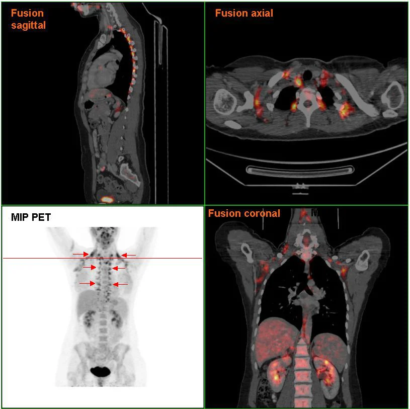
Researchers from Chiba University and partner hospitals found that when mice in a remission‑like state were exposed to restraint stress, the sympathetic nervous system sent signals that bind to β3‑adrenergic receptors on brown adipose tissue (BAT). This stimulation caused BAT to release large amounts of the pro‑inflammatory cytokine interleukin‑6 (IL‑6) into the bloodstream.
From Fat to Joint: The Inflammatory Cascade
The circulating IL‑6 then traveled to joints that had been previously inflamed. There it bound to fibroblast‑like synoviocytes lining the joint capsule, prompting those cells to secrete the chemokine CXCL1. CXCL1 acts as a beacon that attracts neutrophils, a type of white blood cell that inflames the joint, leading to swelling and pain. This chain of events does not rely on the classic autoimmune lymphocytes that usually drive rheumatoid arthritis.
In mice, blocking β3‑adrenergic receptors or the neutrophil chemokine receptor CXCR2 stopped the cascade, preventing stress‑induced joint flares. In humans, a genetic variant that reduces ADRB3 function weakened the link between perceived stress and disease activity in rheumatoid arthritis patients. These findings suggest that targeting the stress‑brown‑fat‑IL‑6 axis could help prevent flare‑ups without broadly suppressing the immune system.