

Brain scans map rapid fear routes between thalamus and amygdala
Scientists have charted several fast‑track routes that connect the thalamus and amygdala, shedding light on how the brain can instantly sense danger.
21 Oct 2026 0 reads Compiled with AI
Researchers used brain scans to identify direct connections between the thalamus, a relay station for sensory signals, and the amygdala, the region that triggers fear responses. These pathways, often called “low‑road” routes, can transmit basic sensory information to the amygdala without first passing through the cerebral cortex. By acting quickly, they help the body prepare for a threat when a sudden noise or sight appears. The study, published in NeuroImage, is the first comprehensive map of these routes in humans.
Mapping the neural routes
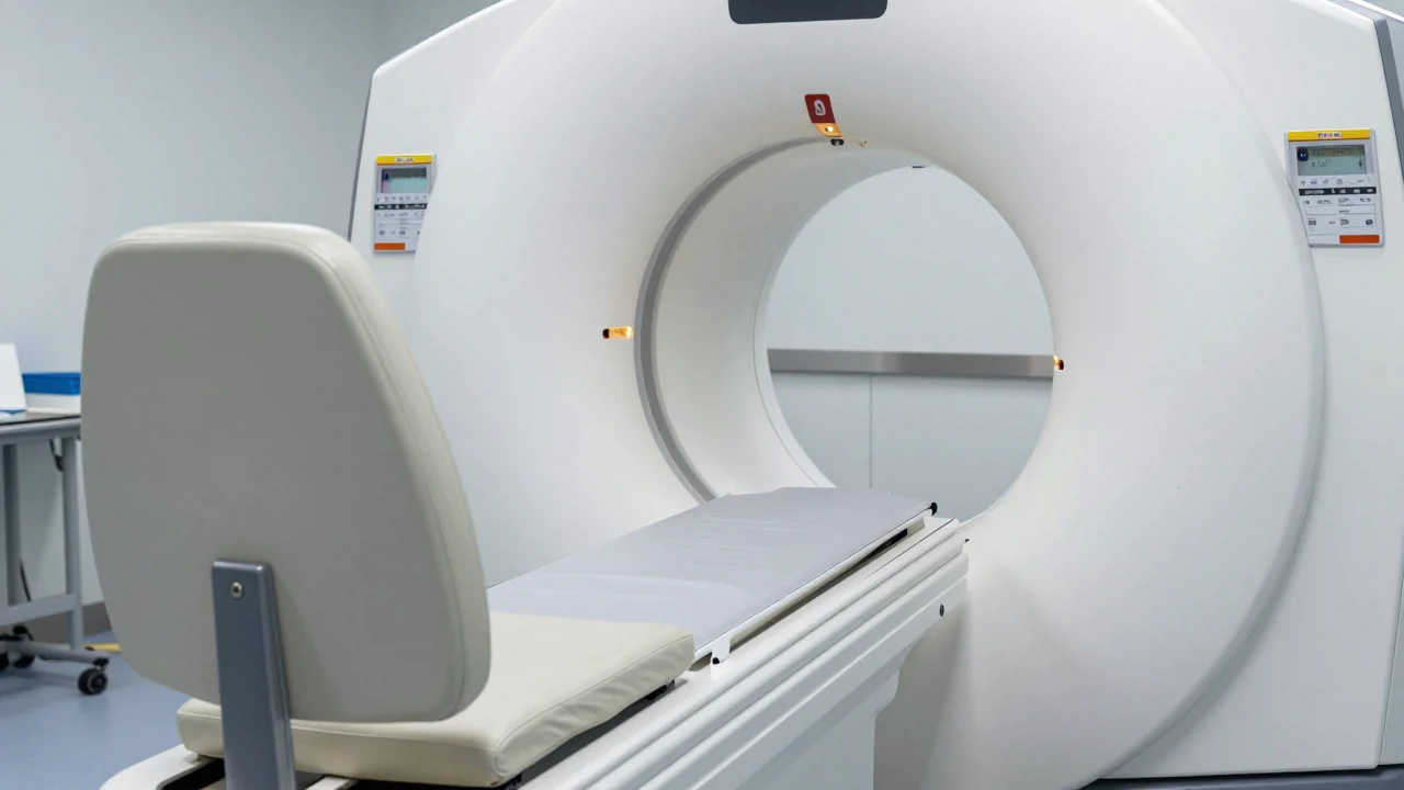
The team examined 113 healthy, right‑handed volunteers with an average age of 24.5 years, including 65 women. They performed high‑resolution diffusion‑weighted MRI scans, which track the movement of water to infer the direction of nerve fibers. Using tractography, they reconstructed probable fiber pathways between several thalamic nuclei and the basolateral amygdala. A subset of 24 participants was scanned again about 15 days later, allowing the researchers to test whether the same connections could be seen across sessions.
The most reliable connections linked the posterior thalamus to the basolateral amygdala. One pathway ran from the inferior colliculus, through the medial geniculate nucleus, to the amygdala, matching an auditory fear pathway known from rodent studies. Connections with the pulvinar, a thalamic region involved in visual attention, were found in all participants and were especially strong for the inferior pulvinar. The mediodorsal nucleus also showed a prominent link, though it may carry signals mainly in the reverse direction. Pathways from intralaminar nuclei varied between people and between the two scans, indicating that some routes are more stable than others.
Key brain connections revealed
These findings show that humans have multiple, rapid thalamus‑amygdala pathways that likely support flexible defensive responses. However, diffusion tractography can sometimes miss real connections or show false ones, especially where nerve fibers cross. The technique also cannot reveal which way signals travel, so the link to fear is inferred from animal work. Future studies that examine brain tissue directly will be needed to confirm these routes and clarify their functional roles.